
Results


#1. What is the diagnosis?
The image illustrates a left VI nerve palsy from herpes zoster ophthalmicus.

#2. What process is illustrated in the radiograph?
The “hair-on-end” appearance of the skull in the radiograph is consistent with massive bone marrow hyperplasia.

#3. The pictured patient suffered a traumatic head injury. What underlying diagnosis is suggested by the examination findings?
Ptosis and mydriasis suggest a cranial nerve III palsy. The appearance of these signs after a crush injury suggests that a skull fracture is impinging on the nerve canal.

#4. This radiograph was taken 12 minutes after infusion of intravenous contrast. What diagnosis is suggested?
The intravenous urogram shows caliectasis and a dilated ureter with columnation (absence of peristalsis), a filling defect around the ureteric meatus, and edema of the distal ureter wall. These findings are most suggestive of a calculus at the right uretero-vesical junction.
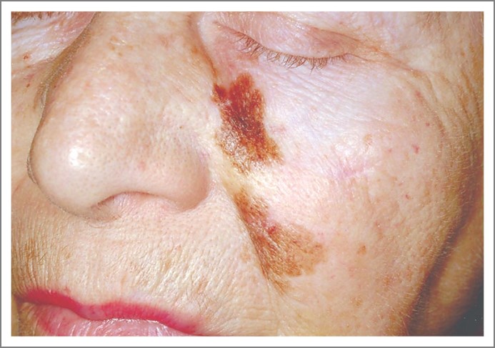
#5. What is the most important diagnosis to exclude in this 81-year-old woman?
The image shows a poorly circumscribed macule with variegated pigmentation. The histopathological diagnosis was lentigo maligna, a diagnosis that should be considered in any pigmented skin lesion.
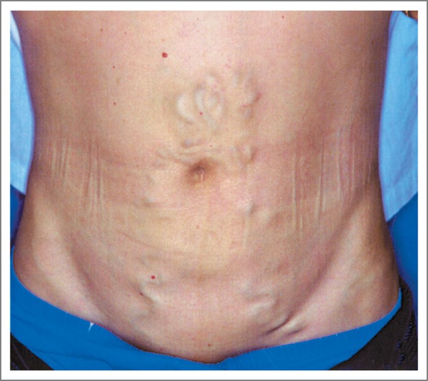
#6. What is the diagnosis?
The image illustrates dilated veins radiating from the umbilicus secondary to portal hypertension.

#7. This 61-year-old man is receiving care for an arrhythmia. What is the cause of his appearance?
The blue-grey appearance is a result of treatment with amiodarone.
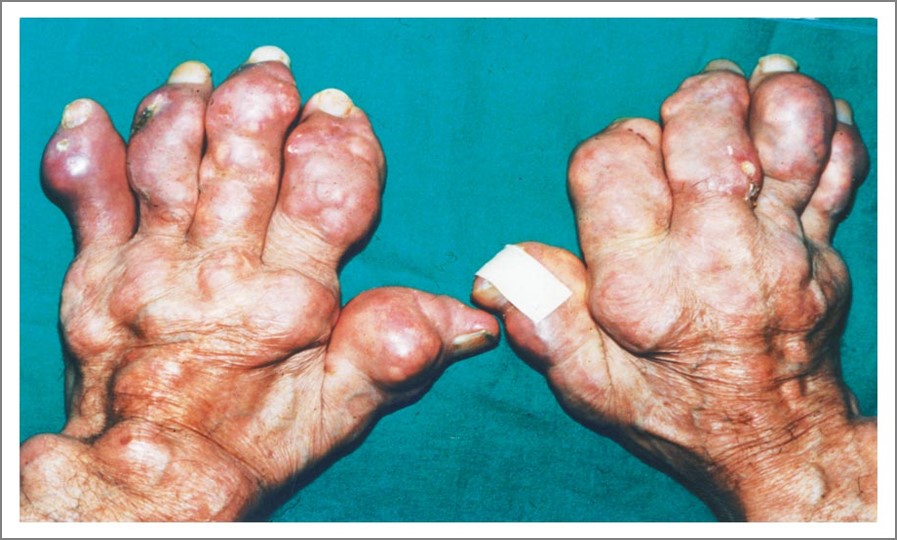
#8. What is the diagnosis?
The image illustrates severe deforming gout.

#9. This urine sample was photographed under light microscopy. What type of crystal is illustrated?
Calcium oxalate crystals.

#10. What is the diagnosis?
The eyeworm illustrated is Loa loa, a filarial parasite endemic to west and central Africa.


